PLAGIOCEPHALY


Introduction:
During recent years, coincident with the recommendation to position infants supine ("Back to Sleep") (7).The "Back to Sleep" campaign has dramatically decreased the incidence of sudden infant death syndrome; however, its sequelae of deformational plagiocephaly have today reached epidemic proportions (8). One in 300 new born is suffering this condition. Positional plagiocephaly has become an increasing problem for pediatricians and craniofacial specialists. Diagnosis is commonly based on history and clinical features, but may be difficult in some cases when characteristic features are missing and radiographic studies seem to be necessary (5).
What is plagiocephaly?
The term plagiocephaly, from the Greek plagios (oblique) and kephalê (head), means distortion of the head, and refers clinically to cranial asymmetry (4). Plagiocephaly is a condition characterized by an asymmetrical distortion (flattening of one side) of the skull. It is a common finding at birth and may be the result of a restrictive intrauterine environment (1).
2 broad causes of plagiocephaly:
According to Margulis A it is a deformity produced by intrauterine and/or postnatal deformational forces. Categorization and diagnosis of plagiocephaly as synostotic or deformational (nonsynostotic plagiocephaly- NSP) is reliably made by physical examination and computerized tomography (2). If there is premature union of skull bones, this is more properly called craniosynostosis. The unusual head shape in plagiocephaly is caused by pressure in the womb giving a "diamond" shaped head when seen from above. In pronounced cases there may be flattening of one side of the chest as well (2).
Torticollis leading to plagiocephaly in newborns:
According to Stellwagen L et al asymmetries of the head and neck are very common in normal newborns. In their study 16% of 102 study newborns were found to have torticollis. Such newborns, especially if they sleep supine, are thought to be at risk of developing deformational posterior plagiocephaly (6).
Identification of affected infants may allow early implementation of positioning recommendations or physical therapy to prevent the secondary craniofacial deformations that are part of an increasingly common phenomenon (6).
Why differential diagnosis in Plagiocephaly in children is important?
In the last decade, the medical fraternity has learned to distinguish deformational plagiocephaly clinically from craniosynostosis (8). Its differential diagnosis is extremely important because prompt surgical correction is usually indicated for the synostotic type. In contrast, infants with deformational frontal or occipital plagiocephaly generally respond to helmet treatment (2). Awareness of deformational plagiocephaly allows more accurate diagnosis and appropriate treatment, avoiding unnecessary surgical intervention in patients with positional molding.
Palpatory diagnosis of plagiocephaly (4).
Sergueef N et al reviewed the mechanics of the occipital bone and the adjacent atlas and bones of the cranial base, in relation to records of 649 functional plagiocephaly children. The review of available data consisted of
Gender, age at presentation, birth history, obstetrical data (breech presentation, vacuum extraction, forceps delivery or Caesarean section), presenting complaint, side of posterior plagiocephaly, side of frontal plagiocephaly, torticollis, motion pattern of the occipital bone upon the atlas, and motion pattern of the spheno-occipital synchondrosis.
Sergueef N et al found a significant correlation between the lateral strain pattern of the spheno-occipital synchondrosis and plagiocephaly and between rotational dysfunction of the occiput upon the atlas and the side of posterior plagiocephaly. Hence they suggested that neonatal osteopathic examination can identify individuals predisposed to develop posterior plagiocephaly.
Advantages of ultrasound in diagnosis of positional plagiocephaly.
Near-field high-frequency ultrasound has been used to evaluate the sonographic findings of suture anatomy and confirm the diagnosis of positional plagiocephaly as well as provide information of prognostic value (5). Regelsberger J et al reported 100 pediatric patients between the ages of 2 and 13 months, who were admitted to their department since 2004 with an abnormal head shape suggesting nonsynostotic plagiocephaly (NSP) diagnosed with High-frequency ultrasound.
They recommend:
High-frequency ultrasound is a relatively inexpensive, safe, and easy-to-use tool for confirming the diagnosis of positional plagiocephaly and excluding true synostosis. Suture anatomy was examined using a 7.5-MHz linear transducer. Morphological characteristics of the sutures--interosseous hypoechoic areas between hyperechoic bone plates--were comparable to those of normal cranial sutures. Overlapping hyperechoic bone plates were found plagiocephaly. Overlapping bone plates may be seen on the affected side of the skull in a majority of plagiocephalic patients, but this finding seems to have no prognostic value regarding early fusion of sutures and therefore should not affect treatment decisions (5).
Because US scan does not involve ionizing radiation, sonography has the potential to be a standard modality for investigating plagiocephaly in infants and should be offered in craniofacial outpatient clinics (5).
Early signs of cranial flattening in healthy neonates (7):
In a study Peitsch WK et al tried to determine whether early signs of cranial flattening could be detected in healthy neonates and to document incidence and potential risk factors.
They proposed that (7):
1. Localized lateral or occipital cranial flattening at birth is a precursor to posterior deformational plagiocephaly. The infant lies supine, with the head turned to the flattened area, and is unable to roll.
2. Intrauterine risk factors for localized cranial flattening are the same as for deformational plagiocephaly.
They concluded (7):
To avoid postnatal progression from a localized cranial flattening to posterior-lateral deformational plagiocephaly, we suggest amending the recommendation of the American Academy of Pediatrics on sleep position: Alternate the head position and allow sleeping on the side and, when awake, supervise prone time.
Role of primary care givers In Plagioceplahy:
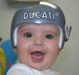
Primary care providers must increasingly be aware of this condition and, in turn, educate new parents about its prevention. Should preventative measures fail and infants develop persistent sleep patterns that result in craniofacial deformities, deformational plagiocephaly can be treated successfully with behavior modification or cranial molding-helmet therapy.
Role of physical therapy & Cranial osteopathy as a complementary treatment of postural plagiocephaly
For the majority of neonates and young infants, appropriate postures and standard physiotherapy succeed in preventing or correcting acquired cranial deformations (fetal due to restricted mobility in utero or postnatal secondary to exclusive dorsal decubitus) (4). Identification of affected infants may allow early implementation of positioning recommendations or physical therapy to prevent the secondary craniofacial deformations that are part of an increasingly common phenomenon (6).
However, Cranial Osteopathy, since it was first proposed, has focussed upon the diagnosis and treatment of birth trauma and cranial asymmetries, and consequently specific therapy for plagiocephalic deformities has been described. Osteopathic manipulation also has been proposed as a treatment for torticollis, a condition associated with plagiocephaly (4).
Why osteopathic treatment is fruitful in plagiocephalic deformities?
1. At first, diagnostic palpation will identify which suture is normally mobile with the respiratory cycle, and which has limited or absent mobility secondary to abnormal postures.
2. Later on, the goal of the therapeutic phase is to mobilise impaired sutures, by various gentle maneuvers depending on the topography of the impairment.
3. The treatment of plagiocephalic deformities in osteopathy is not restricted to the skull but extended to the spine, pelvis and lower extremities which contribute to the deformative sequence.
Osteopathic treatment belongs to complementary medicine, therefore demonstration of its scientific value and favorable results have to be provided. However, referring pediatricians should be more aware of the method and expectations: major deformative sequence since birth and increasing deformations despite preventive postures and standard physiotherapy are reasonable indications for such complementary treatment. Moreover osteopathy has no place in the treatment of craniosynostosis ; the latter belong to malformations, completely distinct from postural deformations (4).
Reference (s):
1. http://en.wikipedia.org/wiki/Plagiocephaly
2. Margulis A; Harefuah. 1999 Apr 2;136(7):532-7, 588, 587.
3. Arch Pediatr. 2008 Jun;15 Suppl 1:S24-30.
4. Sergueef N et al; Complement Ther Clin Pract. 2006 May;12(2):101-10. Epub 2006 Mar 29.
5. Regelsberger J et al; J Neurosurg. 2006 Nov;105(5 Suppl):413-7.
6. Arch Dis Child. 2008 Oct; 93(10):827-31. Epub 2008 Apr 1.
7. Peitsch WK et al ; Pediatrics. 2002 Dec;110(6):e72.
8. Losee JE et al; Clin Plast Surg. 2005 Jan;32(1):53-64, viii.


Comments
Post a Comment