Posts

Showing posts from December, 2009

The Scalene Muscle & Potential pain generating factors from scalene

Image
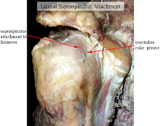
The scalene muscles are a group of three pairs of muscles in the lateral neck, namely the scalenus anterior, scalenus medius, and scalenus posterior. They are innervated by the spinal nerves C3-C8. Origin and insertion Scalenus anterior & medius: Scalenus anterior muscle arises from anterior tubercles of transverse processes of C3-6 vertebrae. It gets inserted into scalene tubercle on the first rib. Scalenus medius arises from posterior tubercles of transverse processes of C2-7 vertebrae. It gets inserted on the superior surface of the first rib behind the groove for subclavian artery. (J.Anat.Soc. India 55 (2) 52-55 2006 52) Function On neck: All 3 scalene muscles produce rotation of the cervical spine to the same side. Maximum stretching of the scalenes should include rotation to the opposite side. (J Orthop Sports Phys Ther. 2002 Oct;32(10):488-96.) On ribcage & respiration: The action of the anterior and middle scalene muscles is to elevate the first rib and rotate the nec

Coupling mechanisms in cervical spine

Image
Radiographic study: coupling of cervical motions in lateral head translations. The clinically common posture of lateral head translation results in an S-shaped cervical spine and may occur in side impact trauma. This posture has not been studied for cervical coupling patterns or range of motion (ROM) (2). In a radiographic experimental study (2) on 20 subjects lateral (a mean of 51 mm) translation was produced by translating the head over a fixed thorax. The major coupled motion was lateral bending (z-axis rotation). The following was found: 1. S-shape in cervical spine was created & there is change in direction at C4–C5 disc space. 2. Upper cervical (C3–C4) lateral bending was contralateral to the main motion of head translation direction. Lower cervical and upper thoracic lateral bending was ipsilateral. 3. During the translation other segmental motions averaged less than 1 mm and 1°. This study shows in cervical spine side bending usually there in nil-minimal involvemen

Effect of eye position on neck muscle activity

Image
Dominant eye assessment has been an integral part of spine conditions especially to reason out the spinal asymmetry. Activity of splenius capitis (SC) is modified with gaze shift. Further, control of the neck muscles is coordinated with the sensory organs of vision, hearing and balance. Impact of eye movement on cervical muscles & movements are not much studied in the context of management of cervical dysfunctions. we can pick up some clues from the study below: The following is a study of division of physiotherapy (University of Queensland, Australia) which investigates interaction between eye movement and neck muscle activity in influencing the control of neck movement. The effect of eye position on neck muscle activity during cervical rotation was studied on 11 subjects. Muscles to rt. Side cervical rotation (in sitting) was under real-time EMG surveillance i.e. right obliquus capitis inferior (OI), multifides (MF), and splenius capitis (SC), and left sternocleidomastoid (SCM).

Rotator cuff tendons & Biceps target area palpation

Image
1. Supraspinatus: Performance cues- 1. Sitting: HBB (Hand behind back)- Adduction & medial rotation of the arm brings the tendon from frontal plane to sagittal plane. 2. Tendon emerge under the anterior edge of acromion 2. Infraspinatus: Performance cues- 1. Pr. Lying: prop up the patients on the elbows 2. Lateral rotation of the shoulder- ask the patient to hold to sides of the couch 3. Adduct the shoulder by shifting the affecting the elbow for 2-3 inches Locating the tendon- The insertion of Infraspinatus tendon is just below the lateral end of the spine of scapula 3. Subscapularis: Performance cues- 1. Sitting or ½ Lying: Therapist palpates for Bacipital tendon by repeatedly IR & ER of the arm. Just medial to the proximal tendon there is lesser tuberosity & the subscapularis (tendon feels as hard as the bone). The tendon extends down to the shaft of humerus distally. 2. Put patient’s hand on her thigh & apply the massage to specific points 3. Caution: Avoid

Dr Alsears letter to me- From medicine to meditation

Image
From medicine to meditation This entire article is by Dr Al Sears. This letter was received by me on my E-mail address, which made a fantastic reading for me. Al Sears, MD 11903 Southern Blvd., Ste. 208 Royal Palm Beach, FL 33411 December 11, 2009 Dear satyajit, When I was in India I met “yogis” or meditation masters who had remarkable powers of concentration. Simply by focusing on their breath they could change their heart rate, raise their body temperature, and even walk on burning-hot coals. In your daily life you probably don’t have the need or opportunity to walk on hot coals, but the power to quiet your mind gives you more control over your health than any pill or prescription. Many of these yogis practice meditation. It’s easy enough to do on your own. There are even clinical studies that back it up. Studies show meditation: * Lowers blood pressure * Improves sleep * Reduces stress and anxiety * Improves immunity * Helps with pain management * Increases produc

Issues in Academics: Writing a PhD Synopsis/Summary/Proposal

Issues in Academics: Writing a PhD Synopsis/Summary/Proposal

Effect of verbal cues on exercise performance

Image
Muscle timing and activation amplitude and movement can be modified with verbal cues (Lewis & Sahrmann ,2009). Lewis & Sahrmann tested the participants under 3 conditions: no cues, cues to contract the gluteal muscles, and cues to contract the hamstrings muscles. Hip and knee angle and electromyographic data from the gluteus maximus, medial hamstrings, and lateral hamstrings was measured while participants performed prone hip extension from 30 degrees of hip flexion to neutral. (see the figure above) This information is important for clinicians using prone hip extension as either an evaluation tool or a rehabilitation exercise.

Pregnancy changes them all- Stability, continence and breathing. The role of fascia in these changes.

Image
(Figure 1) Pregnancy & pelvic Girdle Pain (PGP): Pregnancy-related pelvic girdle pain (PRPGP) has a prevalence of approximately 45% during pregnancy and 20-25% in the early postpartum period. Most women become pain free in the first 12 weeks after delivery, however, 5-7% do not. Pregnancy & urinary incontinence (UI): Wilson, Herbison, Glazener, McGee & MacArthur (2002): found that 45% of women experienced UI at 7 years postpartum and that 27% who were initially incontinent in the early postpartum period regained continence, while 31% who were continent became incontinent. The abdominal canister (container) in pregnancy and delivery: Muscles and fascia of the lumbopelvic region play a significant role in a. musculoskeletal function b. continence and c. respiration It seems that there is an existence of an interlinked trio of lumbopelvic pain, incontinence and breathing disorders. Musculoskeletal loading & the interlinked trio: (see figure 1 above) Synergistic f

Therapy with amitriptyline or physiotherapy is equally effective in fibromyalgia!!!

Image
You can learn about fibromyalgia from this blog site by typing “fibromyalgia” in the search box of this blog site. The search box is at top left of this page. In an rural set up Indian study, first of it’s kind, comparing the effect of amitriptyline or physiotherapy in improving outcome in patients of fibromyalgia over a period of six months, Joshi et al found they are equally effective. Usually fibromyalgia is treated by: antidepressants, analgesics, exercise, cognitive behavioral therapy. 1. Therapeutic Drugs used in fibromyalgia: amitryptiline and other tricyclic antidepressants, pregabalin, duloxetine and milnacipran. 2. Physiotherapy in fibromyalgia: Exercise in the form of cardiovascular training, strength training, aerobics, flexibility training, all can lead to improvement in fibromyalgia. Joshi employed a trained physiotherapist to conduct a uniform structured physical training and aerobic session for patients in physiotherapy treatment group. The patients were advised to perf