Posts

Showing posts from February, 2009

Aggravating activities in chronic low back ache are not specifically directional !!!

Classifiable self reported aggravating activities in chronic low back ache are: aggravation in flexion, extension or unilateral bending. Wand BM et al tried to find do the self-reported aggravating activities of people with chronic non-specific low back pain move the spine in a consistent direction? In an observational study they found there is no evidence for the existence of a consistent direction of spinal movement during the self-reported aggravating activities of people with chronic non-specific low back pain . Participants in this study were described as demonstrating a directional pattern if all three self-reported aggravating activities moved the spine in the same direction. Result of the study by Wand BM et al: In their study over 148 participants with three classifiable aggravating activities they found: 1. Only 47 (32%) demonstrated a directional pattern. And out of them 2. 46 (98%) demonstrated a flexion pattern and 3. 1 (2%) an extension pattern. They have also

SPINE MOBILIZATION CAN AFFECT ANS

Image
See the sagittal image downloaded from google image: http://www.medi-fax.com/atlas/normalspine/images/norm3MRI7.jpg My comments on the image: 1. The slice taken is passing through mid way between two vertebral bodies i.e. passing through the middle of the transverse foramina. 2. look at the inverted triangle- that is the TS of spinal cord. 3. look at the two arms of the triagle- they are the two spinal roots. 4. look at the angle of the triangle from which the roots are coming you can see black dots on a white back ground. That portion is the area that shows TS of roots that are going to exit on levels below. Mixed nerves carrying all fibers i.e. sensory, motor & autonomic fibers can be entrapped here by posterior or posterior-lateral disc prolapses. Automomic fibers control many visceral functions. At lumbar level they control functions of bowel & bladder. Autonomic inputs to control peripheral vascular tone in the lower limb also pass through the lumbar spine. The

How clinical tests are tested:

The clinical tests are tested on the following criteria: 1. Generally accepted criteria for reproducibility (inter- and intra-observer reliability and agreement). 2. Construct validity. 3. Responsiveness. 4. Feasibility.

5-cervical clinimetric tests:

1. Muscle endurance of short neck flexors This test was first described by Grimmer, and several modifications have been described since then. Test position: The patient in supine position. The modified test position is crook lying. Subjects are instructed to "tuck in their chins" (craniocervical flexion) and then to raise their heads. The time between assuming the test position until the chin begins to thrust is measured in seconds with a stopwatch. Modification of the original test by Grimmer: In these modifications, the starting position for the test is different (crook lying) and the examiners monitor the chin tuck and occipital position. 2. Manual muscle testing Test position: Test is performed without head support, prone for extensors and supine for flexors. Manual resistance is applied and strength is graded. Grade 1= enable to maintain position against gravity. …….. Grade 5= maintaining position against full manual resistance. 3. Craniocervical flexion test Patient pos

Clinimetric evaluation of methods to measure muscle functioning in patients with non-specific neck pain

Image
Panjabi et al estimated that the neck musculature contributes about 80% to the mechanical stability of the cervical spine, while the osseoligamentous system contributes the remaining 20%. Evidences suggest that patients with neck pain have 1. Reduced maximal isometric neck strength and endurance capacity. 2. Further in patients with chronic neck pain jerky and irregular cervical movements and poor position sense acuity have been found. Exercises are commonly used to improve neck muscle function and thereby decrease pain or other symptoms. Evaluating the progress of neck muscle function during treatment requires tests which can be carried out easily and meet certain standards for clinimetric properties. Chantal HP de Koning et al of Netherlands did a review to provide information for researchers and clinicians to facilitate choices amongst existing instruments to measure neck muscle functioning in patients with neck pain. Following are their conclusions: 1. The choice of a test

Frozen Shoulder Update

Image
Various stretches have been introduced for the posterior shoulder; however, little quantitative analysis to measure stretching of the posterior capsule has been performed. Which further implies the biomechanical impacts of the stretching on the joints would vary. That further implies that the out comes on joint mobility of a stiff joint are different. In clinical setting we want quick and more importantly less painful techniques that are easy to administer by the therapist. I personally have utilized the cyriax’s capsular stretching methods for a decade now. I am currently working on modified techniques of my own. To improve the hand behind the back my personal favorite is mulligan’s technique. However I have modified that too in my own way. Currently I am treating 7-8 frozen shoulders a day in my clinic. Personal satisfaction is what I cherish most. But with satisfaction there comes dissatisfaction; I hope that’s true for any clinician who understands his patient’s plethora of sufferi

rotator cuff update

Image
An investigation of supraspinatus tendon samples obtained from patients undergoing arthroscopic repair of a rotator cuff tear revealed: 1. Cartilage-like changes in rotator cuff. According to the researchers stress-shielded and transversely-compressed side of the enthesis of the rotator cuff has a distinct tendency to develop cartilage-like or atrophic changes in response to the lack of tensile load. The formation of cartilage-like changes in the enthesis in many ways can be considered a physiological adaptation to the compressive loads. 2. Rotator cuff tendons also show frequently marked changes on articular side of the rotator cuff under arthroscopic vision. 3. Over a long period, this process (described above) may develop into a primary degenerative lesion in that area of the tendon. 4. According to the authors these above 3 points explains well why the tendinopathy is not always clearly activity related, and can be strongly correlated with age. 5. The authors due to above said reas

Fatness affects the overall oxygen delivery to working tissues but not fitness

Image
I have grown up as a sports physiotherapist reading clinical sports medicine by Brukner & Khan. Till now this book is sort of bible to me. i am fascinated by the book and more by the author's ability to express terse subjects in explicit yet digestible manner. when i saw a article by Karim Khan in British journal of sports medicine, i was quite eager to know what is it all about. it was fitness of fat people. Few important points are as follows: 1. changes in fitness & changes in fatness may not go side by side: Aerobic fitness is the overall ability of the cardio-respiratory system to transport litres of oxygen to the working tissues. Any accumulation of body fat reduces the utility of this transport in terms of daily activities (including treadmill running). Example: To take a practical example, a man with a body mass of 70 kg and an aerobic fitness of 3.5 l/min has a relative VO2max of 50ml/[kg.min]. If that same person accumulates an extra 14 kg of body fat, the aerobi

The way I treat my Bell's Palsy cases

Image
Bells palsy is a LMN type of lesion of facial nerve. usually theere is a compressive pathology i.e. a oedema withing the outlet of facial nerve in the mastoid canal. the muscles of one side of the face are flaccid. In many of my patients i found the following history: Travel in bus or car for a long journey, with cold air hitting the affeted side face. i few cases patients repot of taking icecream & ear-ache. some common suspected causes are: - virus - compression of nerve - bacteria - drugs - cold temperature - trauma of the facial nerve - Pressure of facial nerve due to tumor As a physical therapist i recommend a 21 day physical therapy course consising of following treatment modalities (i dont how many of these modalities are evidence based but i have been using them for at least 10 years now): 1. Facial Massage for 5-10 minutes twice a day in a chin - forehead direction to maintain the tone. 2. Infrared on the affected side to increase the blood supply and decrease skin resista

A Simple Review of Shin Splint

Image
MTSS is the short form of medial tibial stress syndrome which is an advanced medical terminology used to describe a group of exercise induced leg pain. Shin splint is one of the exercise induced leg pain. In the following article we will discuss about shin splints. Ways shin splint presents: 1. Compartment Syndrome: Where an increase in pressure in certain muscles can cause extreme pain and decreases in circulation to the leg. 2. Tendonitis/Periostitis: Abnormal strain placed on the muscles, tendons, and on the covering around the bone that the tendon is attached to. 3. Stress Fractures: When exposed to increased strain and fatigue, minute fractures may result in the bones of the lower leg. Contributing factors: • Poor walk/run mechanics • Inadequate calf flexibility • Inadequate strength of the dorsi-flexors (muscles of the front of the lower leg) • A rapid increase in mileage or frequency of aerobic classes • Poor or worn out footwear Symptoms include: • Pain – usually on the insi

The place of Physioanatomic Imaging Approach in treatment of LS spine disorders:

Image
The following should be an algorithmic approach of clinicians working with LS spine disorders. 1. Patient symptoms and history must be evaluated by use of a pain drawing and information sheet. 2. The patient's pain pattern is categorized into a nonspecific pattern or into one of four recognizable pathway patterns (radicular, dorsal ramus, polyneuropathy, and sympathetic). Because each spinal lesion is typically manifested primarily via one of the four symptom pathways, the distribution of expected symptoms from each pathologic feature can be compared with the patient's pain drawing. 3. The patient's presenting symptoms are also used to determine the most cost-effective and efficacious use of initial diagnostic imaging evaluation. 4. In a minority of patients the findings on noninvasive imaging either will not correlate with the patient's symptoms or will demonstrate multiple abnormalities that could account for the patient's symptoms. In these patients,

will you succeed in treating a cervical redicuopathy?

Image
while a patent is under your care (physiotherapy) you can know with certain conviction whether the patient is going to improve or not. variable for the prediction: According to Cleland, J.A et al the following cluster of variables as most significant: Age of less than 54 years Dominant arm not affected Looking down does not worsen symptoms Multimodal treatment (manual therapy, cervical traction, deep neck flexor strengthening for at least 50% of visits) Predictions: 1. Having three of the four variables present led to an 85% posttest probability the patient would experience short-term success. 2. Having all four variables present were associated with a 90% post-test probability .

8- Clinical tests for testing rotator cuff tears

3 most encountered rotator cuff pathology are: bursitis, partial-thickness rotator cuff tear, and full-thickness rotator cuff tear. The following eight clinical tests were examined: * Neer impingement sign * Hawkins-Kennedy impingement sign * Painful-arc sign * Supraspinatus muscle strength test * Infraspinatus muscle strength test * Speed test * Cross-body adduction sign * Drop-arm sign watch the following site for details: http://www.aafp.org/afp/20000515/3079.html

Sports Shoes. Elementary considerations of sports shoes: part I

Image
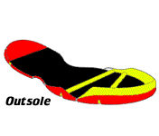
Different sports have different shoes. For example horse riding & ballet dance shoes are different that of running or football shoes. This following topic is written with reference to running & sports requiring similar shoes. Athletic running sports shoes bring automatically an idea that they are soft on feet & absorb shock. However, Withnall R et al studying on army recruits reported similar rates of lower limb injuries were for all insoles (shock absorbing and non-shock absorbing) in a randomized controlled trial. The trial provides no support for a change in policy to the use of shock absorbing insoles for military recruits (4). To clarify this ambiguity we found an opportunity to write an article is to assist in understanding athletic footwear. In addition, the various components of a typical athletic shoe are described, including the upper, the midsole/outsole. Hence let us review the parts of a sports shoe. Parts of a sports shoe: 1. Upper 2. Insert 3. Midsol

Sports Shoes. Prominent injuries by sports shoes (Evidences only): part II

Though improper sports shoes could have a direct relationship with sports injuries, there are not many direct evidences that link sports shoes to a particular kind of sports injuries. We analyzed the recent evidences from the year 1994-2008 from PUBMED with the key word “sports shoes” AND “sports injuries”. In a recent research it was found no increased incidence of ankle sprains is associated with shoe design in collegiate basketball players (2). How ever this finding is hard to believe in every sporting scenario. Taunton JE et al found running shoe age were associated with injury. Knee was the most injured joint found in the study however the sports shoes are not incriminated in all cases of injuries(3). Similarly according to Milburn PD et al literature related to footwear design and injury prevention in most sports played on natural turf is limited (5). Following are few direct evidence we found in above said search criteria: According to Van Mechelen there are many associations be

Sports Shoes. Protective aspect of sports shoes & current concepts: part III

1. The protective function of sports shoes. The shoe can be thought of as a powerful tool for controlling human movement. A well-designed shoe can assist in reducing the number of lower limb injuries arising from sport and training activities (9). Barnes RA et al reviewed the types of injury acquired by sportsmen in both training and playing is then followed by a discussion of aspects of footwear design and their role in both contributing to and preventing lower limb injury. Finally, the paper considers support and shock absorption techniques in the context of footwear design (9). Working on hip injuries Paluska recommends; coaches, trainers and medical personal who care for runners should advocate running regimens, surfaces, shoes, technique and individualised conditioning programmes that minimise the risk of initial or recurrent hip injuries (5). Running shoes are designed specifically for different foot types in order to reduce injuries. Running in the correct footwear matched for f

Review of Labral Tears of shoulder

Image
The glenoid labrum of the shoulder has extensive anatomic variation but appears to be important for contributing to shoulder stability and for increasing the depth of contact between the glenoid labrum and the humeral head. Tears of the labrum are commonly seen in association with other pathologic entities, such as instability and rotator cuff tears, and treatment of the labral pathology may be incidental to treatment of the other more significant pathology. However, conditions isolated to the labrum do occur and can be a significant source of shoulder problems. Effective treatment of these lesions may result in significant improvement in the patient's symptoms (4). Labral lesions are difficult to diagnose, and special diagnostic studies and, frequently, arthroscopy are required. How much we can rely on clinical tests is a question. Shoulder complaints are frequently recurrent. Instability might cause some of these complaints (3). History taking and clinical tests are commonly used

Discussion of Mechanisms Of Maual Therapy

Image
Mechanisms of Manual Therapy : This article is taken from an article in Manual Therapy by Joel Bialosky and associates from the University of Florida. The article provides a framework of manual therapy that has yet to be previously defined to this degree. How manual therapy works is more controversial than the procedures. Convictions differ considerably about proposed mechanisms for manual therapy among the clinicians, teachers & peers. However,in generic terms, now a days it is believed that the identification and correction of biomechanical faults within the musculoskeletal system leads to the clinically found effects. The peer reviewed literatures are proving us insights into what really happens when manual therapy is done. However with emerging evidences, it is gradually becoming clearer that manual techniques are more than correcting upslips and stretching joint capsules etc. Bialosky et al has proposed five potential mechanisms reponsible for manual therapy. Mechanical St

BICYCLING REALTED SPORTS INJURIES

Image
The following painful conditions & injuries may happen in bicyclists. The conditions described below are cephalo-caudally (head to toe) on not on prevalence rates. 1. Neck pain 2. Back pain 3. Shoulder pain 4. Wrist pain 5. Handlebar palsy 6. Groin injuries- saddle sore, chafing, pudendal neuropathy, male impotence, female uvula trauma etc 7. Patello-femoral joint pain (knee cap pain) 8. Foot & ankle paresthesia (numbness in foot & ankle) 9. Metatarsalgia (present as toe pain & numbness) 10. Achilles tendonitis (presents as heel cord pain) 11. Planter fasciitis (presents as heel pain) Friends, in the next part we will discuss how these injuries are caused by riding a bicycle.

treatment of CRPS

Image
How is Complex Regional Pain Syndrome Treated? Because there is no cure for complex regional pain syndrome, treatment is aimed at relieving painful symptoms so that people can resume their normal lives. The following therapies are often used: • Physical therapy: A gradually increasing physical therapy or exercise program to keep the painful limb or body part moving may help restore some range of motion and function. • Psychotherapy: Complex regional pain syndrome often has profound psychological effects on people and their families. Those with complex regional pain syndrome may suffer from depression, anxiety, or post-traumatic stress disorder, all of which heighten the perception of pain and make rehabilitation efforts more difficult. • Sympathetic nerve block: Some patients will get significant pain relief from sympathetic nerve blocks. Sympathetic blocks can be done in a variety of ways. One technique involves intravenous administration of phentolamine, a drug that blocks sympath

Complex Regional Pain Syndrome- CRPS

Image
What is Complex Regional Pain Syndrome (CRPS)? Complex regional pain syndrome (CRPS) is a chronic pain condition that is believed to be the result of dysfunction in the central or peripheral nervous systems. Older terms used to describe complex regional pain syndrome are: • reflex sympathetic dystrophy syndrome (RSDS) • causalgia Causalgia was a term first used during the Civil War to describe the intense, hot pain felt by some veterans long after their wounds had healed. Typical features of complex regional pain syndrome include dramatic changes in the color and temperature of the skin over the affected limb or body part, accompanied by: • intense burning pain • skin sensitivity • sweating • swelling CRPS I is frequently triggered by tissue injury; the term describes all patients with the above symptoms but with no underlying nerve injury. Patients with CRPS II experience the same symptoms but their cases are clearly associated with a nerve injury. Complex regional

An Manual therapy prospective of the Occipitoatlantal Joint

Image
Occipitoatlantal (OA) joint is the most superior weight-bearing synovial joint in the body. Because the OA articulation is one of the final locations at which the body can adapt to asymmetry or dysfunction below, hence this joint requires evaluation in the context of the entire body. The Atlanto-occipital joint (articulation between the atlas and the occipital bone) consists of a pair of condyloid joints.The ligaments connecting the bones are: * Two Articular capsules * Posterior atlantoöccipital membrane * Anterior atlantoöccipital membrane * Lateral atlantoöccipital The movements permitted in this joint are: * (a) flexion and extension, which give rise to the ordinary forward and backward nodding of the head. * (b) slight lateral motion to one or other side. Flexion is produced mainly by the action of the Longi capitis and Recti capitis anteriores; extension by the Recti capitis posteriores major and minor, the Obliquus superior, the Semispinalis capitis, Splenius capitis, Sternocle

Treatment Hyiod Dysfunctions

Image
Introduction: The hyoid bone and related myofascial connections of the neck often are not evaluated as possible causes of head and neck pain or dysfunction. Hyoid bone syndrome involves nonspecific cervical pain and pain while swallowing, with radiating pain to the face, neck and shoulders. This article provides information on hyoid dysfunction, anatomy, evaluation and manual treatment methods. Examination: The working understanding of the anatomy of the anterior structures of the neck is necessary to evaluate patients for this syndrome (a review of that anatomy is provided). Patients with anterior neck symptoms or tension and reduced extension of the neck should be checked for hyoid dysfunction, using palpation on the supine patient. Once the hyoid is located, it is tested for mobility, asymmetry, discomfort and muscle tenderness in the anterior cervical triangles. Bimanual palpation of the suprahyoid muscles is also performed. Treatment: The author discusses five common treatment

Rotator cuff muscles that causes shoulder pain

(1) Supraspinatus: This is one of the three rotator cuff muscles infamously known for giving rise to shoulder pain. It arises from the fossa above the spine of the shoulder blade bone known as supraspinous fossa. It inserts into the upper facet of the greater tuberosity of the arm bone and from the capsule of the shoulder joint. It assists the deltoid in moving the arm away from the side of the body (abduction). It also helps to roll the arm outward (external rotation). It is supplied by the suprascapular nerve from the upper trunk of the brachial plexus. The suprascapular nerve carries the C5 and C6 nerve nerve root fibers (especially the C5 nerve root fibers) to this muscle. The tendon of this muscle is commonly involved in degenerative processes and may rupture. It can also be entrapped under the acromion. (2) Infraspinatus: Shoulder pain symptoms are commonly associated with a rotator cuff problem. The infraspinatus is the largest of the three muscles comprising the rotator cuff,

Internal rotation resistance strength test (IRRST)

One study introduces a new sign to differentiate between outlet impingement and non-outlet (intra-articular) causes of shoulder pain in patients with positive impingement sign: the internal rotation resistance strength test (IRRST). It was hypothesized that positive test results are predictive of non-outlet impingement, whereas negative test results confirm outlet impingement. A prospective comparison between IRRST and arthroscopic findings of 115 consecutive patients showed the test to be highly accurate in differentiating between these two diagnoses (positive predictive value 88%, negative predictive value 96%, sensitivity 88%, specificity 96%, and accuracy 94.5%). The IRRST, in conjunction with impingement and apprehension signs, adds to our armamentarium of tests that distinguish between subacromial outlet impingement and intra-articular forms of pathology. source: (J Shoulder Elbow Surg 2001;10:23-7.)

Clinical test for DD of Labral tears and acromioclavicular joint abnormalities

Labral tears and acromioclavicular joint abnormalities were differentiated on physical examination using a new diagnostic test. Step 1. The standing patient forward flexed the arm to 90° with the elbow in full extension and then adducted the arm 10° to 15° medial to the sagittal plane of the body and internally rotated it so that the thumb pointed downward. Step 2. The examiner, standing behind the patient, applied a uniform downward force to the arm. With the arm in the same position, the palm was then fully supinated and the maneuver was repeated. The test was considered positive if pain was elicited during the first maneuver, and was reduced or eliminated with the second. Pain localized to the acromioclavicular joint or “on top” was diagnostic of acromioclavicular joint abnormality, whereas pain or painful clicking described as “inside” the shoulder was considered indicative of labral abnormality. Reference: Stephen J. O’Brien et al

Diagnostic implications of CSF in Lumbar Disc Herniation

1. Cerebrospinal fluid protein concentrations in patients with sciatica caused by lumbar disc herniation. The increase of the CSF total protein concentration in sciatica without spinal block is assumed to be due to leak of plasma proteins into the CSF from the nerve root. A relationship between CSF protein concentrations and certain clinical parameters has been found. Skouen JS et al studied 180 adult patients admitted to the Neurological Department, Haukeland Hospital in Bergen, Norway, for a period of 5 years from 1984 to 1988. One hundred fifty-seven patients were followed up 3.9-9.0 years after admittance to the Neurological Department. The purpose of this study was to find out if the total cerebrospinal fluid (CSF) protein concentration could predict the outcome of lumbar disc surgery or conservative treatment in patients with sciatica. Elevated CSF total protein concentration was related to chronic leg pain, leg pain, and subjective physical disability at follow-up. Hence the aut

Evidence for shoulder girdle dystonia in selected patients with cervical disc prolapse.

Some patients with cervical disc herniation suffer from persistent nuchal pain and muscle spasms after decompressive surgery despite the lack of clinical and radiological signs for actual spinal root compression. Sonographic examination of the brain in some of these patients showed increased echogenicity of the lentiform nuclei as described in patients with idiopathic dystonia. This has been linked to an altered Menkes protein level and copper metabolism. The authors have suggested a relationship between persistent nuchal pain after adequate cervical disc surgery and dystonic movement disorders. Thirteen patients with persistent nonradicular nuchal pain after at least one cervical disc surgery and without evidence of continuing spinal root compression and 13 age-matched controls were included. All patients had a complete neurological examination, ultrasound, and MRI scan of the brain. In addition, Menkes protein mRNA levels of leucocytes were analyzed in patients and controls. All pat