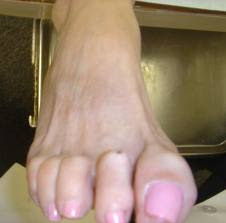
Claw toe deformity

A claw toe is a toe that is contracted at the PIP and DIP joints and can lead to severe pressure and pain. Ligaments and tendons that have tightened cause the toe's joints to curl downwards. Claw toes may occur in any toe, except the big toe. There is often discomfort at the top part of the toe that is rubbing against the shoe and at the end of the toe that is pressed against the bottom of the shoe.
Causes:
Claw toe deformity results from altered anatomy and/or neurologic deficit, resulting in an imbalance between the intrinsic and extrinsic musculature to the toes.
1. Claw toe deformity can develop as a complication of fracture of the tibia. The deformity develops following a tibia fracture is basically due to adhesions of the flexor hallucis longus (FHL) and flexor digitorum longus (FDL) muscles to the surrounding structures under or just proximal to the flexor retinaculum.
According to Fitoussi et al it may be related to a subclinical compartment syndrome localized in the distal part of the deep posterior compartment. Soft-tissue release without tendon lengthening allowed recovery in all patients.
2. Involvement of distal tibial nerve in claw toe. Generally clawing in the foot is related to the lateral plantar nerve involvement but surgeons Dellon & colleagues’ demonstrated improvement in toe clawing may result from neurolysis of the tibial nerve as well as the lateral plantar nerve.
3. Claw toe may develop secondary to diabetic neuropathy affecting foot. Clawing of the toes in the diabetic neuropathic foot is believed to be caused by muscle imbalance resulting from intrinsic muscle atrophy.
According to Bus et al neither intrinsic muscle atrophy nor muscle imbalance causes claw toe deformity in diabetic neuropathy. They also suggested that the role of these muscle factors in claw toe development may not be primary. Their research also suggested a complex nature of development, potentially involving anatomical and physiological predisposing factors.
Claw toe of relatively rapid progression is possibly due to neural involvement. Consult a neurologist as soon as possible.
References:
1. Fitoussi F et al; J Child Orthop. 2009 Oct;3(5):339-43. Epub 2009 Aug 22.
2. Dellon AL et al; Microsurgery. 2008;28(5):303-5.
3. Bus SA et al; Diabetes Care. 2009 Jun;32(6):1063-7. Epub 2009 Mar 11.


Comments
Post a Comment