Posts

Implication of anterior drawer test, Lachman’s test, Pivot shift test to that of knee Instability

Image
Lachman's vs. Anterior Draw Test •    Lachman's test may be more difficult for clinicians to perform but tends to be more sensitive •    In the anterior draw test knee is positioned so that the hamstrings have a mechanical advantage. Increased hamstring activity can inhibit tibial translation, causing a false negative test •    A torn meniscus can act as a block to tibial motion, again causing a false negative while doing the anterior draw test Anterior drawer test with tibia external rotation: Anterior drawer test with tibia in neutral rotation demonstrates equal displacement of both condyles & this displacement is eliminated by internal rotation of the tibia, then both anteromedial and anterolateral rotary instability may be present. Similarly positive anterior drawer test in neutral tibial rotation, that is accentuated when the test is repeated in 30 deg of external rotation and reduced when performed with the tibia in...
C6-C7 syndrome: My clinical experience & suggestions

Image
Many confuse marked tenderness at the coracoid tip, lateral pectoral and medial elbow sites to be a variant of fibromyalgia however usually it comes along with tenderness at the C6-7 level in the cervical spine. A close look in to the history reveals in this group of patients lose tenderness at C5-6 and standard upper body sites with proper neck support during sleep, but remained symptomatic at coracoid tip, lateral pectoral, medial elbow and C6-C7. Smythe HA (1994) have supported that mechanical factors determine patterns of symptoms and tenderness in this group. This implies that we are talking of segmental referred pain or referred tenderness rather than a pathologically ill-defined spectrum called “fibromyalgia”. If we differ on this front our treatment strategy is mislead. That’s why centrally acting medications or behavioral modifications are equally disappointing outcomes. To add to that tricyclic medications, stretch and spray or trigger point injections may be simply ...

Stiff man syndrome: A neuromotor disorder- Know how for physios

Synonyms: stiff man syndrome (SMS), stiff person syndrome (SPS), stiff leg syndrome (a focal SMS) etc Introduction & definition: Moerch and Woltman reported the first 14 cases with this syndrome for more than 50 years since 2010. Stiff man syndrome (SMS) is an uncommon (rare) neurological disease that manifests with disorder of motor function which is characterized by rigidity of axial musculature and fluctuating painful spasms, which are often induced by startle or emotional stimuli. Clinical and immunological findings indicate that SMS is a heterogeneous disease, suggesting the need to define its diagnostic criteria. But it has basic 2 forms either it can be generalized or focal. Criteria for the diagnosis of SPS were proposed but there are several variants of this syndrome described. These variants include focal variants of SMS. Variants include stiff limb syndrome (SLS), jerking SMS and progressive encephalomyelitis with rigidity and myoclonus (PERM). Recognition of the clini...

Soft tissue mobilization by instruments

Image
To spare the treating hands of the clinicians & more to increase the precision of treatment in musculoskeletal therapy, clinicians now days are more and more inclined to use instruments for soft tissue mobilization. None the less these equipments can also be used to diagnose the soft tissue lesions more accurately enhancing the palpation capacity of the clnician. However, the objectives of this form of treatment are as follows: • Break down scar within the tissues • Promote blood flow into the sore area • Establish healing in damaged tissues • Release adhesions within the layers of tissue • Improve lymphatic circulation David Grastron is the pioneer in developing the new area instruments for soft tissue mobilization. However stylus massage was reported to be used in trigger point deactivation by Russian in Olympic game. Spa professionals use various types of massage equipments during spa sessions. Three common treatments always come into mind when soft tissue mobiliz...

GIRD- Glenohumeral internal rotation deficit

Image
Definition of GIRD: GIRD is a 20° or greater loss of internal rotation of the dominant shoulder compared with the non-dominant shoulder. Introduction: Glenohumeral internal rotation deficit, often diagnosed in players of overhead sports, has been associated with the development of secondary shoulder lesions. Conditions such as labral and rotator cuff injuries have been linked with decreases in glenohumeral internal-rotation and increases in external-rotation motion. This group also shows a loss of horizontal or cross-body adduction in the throwing shoulder when compared with the non-throwing shoulder. GIRD is also strongly associated with scapular dyskinesis. Tennis players, swimmers & athletes in throwing sports are commonly affected by GIRD. Deficit in dominant shoulder of tennis players is about twice the deficit found in swimmers. Data suggest that GIRD and scapular position change worsens as the level of competition increases in overhead sports. Pathologic conditions in the sh...

Radiological interpretation of joint space narrowing & Kellgren-Lawrence (K-L) scale

Image
Grade 0 = Normal Grade 1 = Doubtful narrowing of the joint space & possible osteophytic lipping Grade 2 = Definite Osteophytes & definite narrowing of joint space Grade 3 = Moderate multiple Osteophytes, definite narrowing of joint space, some sclerosis & possible deformity of bone contour Grade 4 = Large Osteophytes, marked narrowing of joint space, severe sclerosis & definite deformity of bone contour

Internal impingement of shoulder: A simple overview

Image
The impingement in shoulder can clearly be classified into internal & external varieties. Internal impingement: The internal impingement syndromes result from the impingement of the soft tissues of the rotator cuff and/or joint capsule on the glenoid or between the glenoid and the humerus. External impingement: The external impingement syndromes result from the impingement of the soft tissues of rotator cuff and bursa on the structures of the coracoacromial arch. External shoulder impingement and rotator cuff disease has been corroborative despite of research arguments. Attempts have been made to identify objective imaging criteria that confirm the diagnosis of impingement, but at present external impingement remains primarily a clinical diagnosis. Mainly shoulder impingement is caused by compression of the supraspinatus tendon underneath the coracoacromial arch, mostly in forward flexion of the arm. Stages of external impingement: Different stages of impingement syndrome are descr...

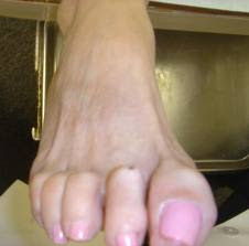
Claw toe deformity

Image
A claw toe is a toe that is contracted at the PIP and DIP joints and can lead to severe pressure and pain. Ligaments and tendons that have tightened cause the toe's joints to curl downwards. Claw toes may occur in any toe, except the big toe. There is often discomfort at the top part of the toe that is rubbing against the shoe and at the end of the toe that is pressed against the bottom of the shoe. Causes: Claw toe deformity results from altered anatomy and/or neurologic deficit, resulting in an imbalance between the intrinsic and extrinsic musculature to the toes. 1. Claw toe deformity can develop as a complication of fracture of the tibia. The deformity develops following a tibia fracture is basically due to adhesions of the flexor hallucis longus (FHL) and flexor digitorum longus (FDL) muscles to the surrounding structures under or just proximal to the flexor retinaculum. According to Fitoussi et al it may be related to a subclinical compartment syndrome localized in the distal...

See how physical therapy specilists are certified in USA

Asian physios please see the following web site to appreciate the job APTA is doing uplifting the physical therapy profession. American board of physical therapy specialists (APTA certifying the physical therapy specialists) http://www.abpts.org/home.aspx

Analysis of sitting posture

Introduction: Occupations & occupational demands have changed in the recent era. Modernization & Industrialization has changed the face of occupational activities & need of work related physical performances. We have slowly crawled in to mostly a sedentary era. On the other side passive leisure time pursuits are taking over active leisure time pursuits. The situation is such that the average office going adult hardly moves his or her axial & apendicular joints in it’s full range of motion. Mean temporal classification of ADL (activity of daily living) closely equates 7-8 hours of sleeping; 8-10 hours of working and rest of hours are spent in house hold activities like washing, watching TV, purchasing grocery, rarely gardening or a sports etc. Office hour activities span more than one third of the day. Except in blue collar jobs (manual labor class) office hours consists of at least 2-4 hours sitting to full office hour invested in sitting like in banking, IT sector job...

Can occupational sitting cause LBA?

Image
Introduction: Sociocultural, economic, and manufacturing factors are 3 different major factors that influence sitting. "Correct" seated posture is associated with spine health was known to people possibly from Hippocratic era. But the debate has not dampened yet whether the sitting is directly a cause of LBA or it predisposes or precipitates LBA. From 1980s much of research has been put into ergonomically correct sitting & providing a work environment for correct sitting posture. However there is a question; is there any medically correct sitting posture? The correct sitting posture refers to maintenance of a correct lumbar spine lordosis actively (muscle contraction) or passively (chair or sitting back support) (described below elaborately in neutral spine position). Passive maintenance is sought because it is noticed that within minutes of sitting lordosis is reversed or lost. According to Ernst even the correct sitting is not out of danger because of the repetitive nat...

Hemiplegia recovery: Newest developments- Abstract from PUBMED

Annu Rev Med. 2009;60:55-68. Stroke rehabilitation: strategies to enhance motor recovery. O'Dell MW, Lin CC, Harrison V. Department of Rehabilitation Medicine, New York-Presbyterian Hospital, Weill Cornell Medical Center, New York, New York 10021, USA. mio2005@med.cornell.edu Abstract Recent evidence indicates that the brain can remodel after stroke, primarily through synaptogenesis. Task-specific and repetitive exercise appear to be key factors in promoting synaptogenesis and are central elements in rehabilitation of motor weakness following stroke. Expert medical management ensures a patient is well enough to participate in rehabilitation with minimal distractions due to pain or depression. Contraint-induced motor therapy and body-weight-supported ambulation are forms of exercise that "force use" of an impaired upper extremity. Technologies now in common use include robotics, functional electrical stimulation, and, to a lesser degree, transcranial magnetic stimulation a...

The SC joint injuries: A mini Review for Physiotherapists in acute care

Image
Parts of SC joints: The sternoclavicular articulation is a double arthrodial joint. The joint is made out of following boby articulating parts sternal end of the clavicle, the upper and lateral part of the manubrium sterni, and the cartilage of the first rib. The articular surface of the clavicle is much larger than that of the sternum, and is invested with a layer of cartilage, which is considerably thicker than that on the latter bone. Important parts (ligaments & disc) of this joint are: 1. The Articular Capsule 2. The Anterior Sternoclavicular ligament 3. The Posterior Sternoclavicular ligament 4. The Interclavicular ligament 5. The Costoclavicular ligament And the 6. The Articular Disk According to a retrospective analysis articular disk injuries were seen in 80% of patients. Injuries of the anterior, posterior, interclavicular and costoclavicular ligaments were seen in 73%, 39%, 29% and 14% of patients, respectively (10). Movements possible in this joint- T...

Autonomy and the future of physiotherapy.

Link: http://www.thefreelibrary.com/Autonomy+and+the+future+of+physiotherapy.-a0181366670 Either follow the link. If the link is not working properly then copy the above link in a web browser. Then type search. I assure you this is a topic to ponder.

Respiratory muscle stretch gymnastics (RMSG) a review of PUBMED from 1996-2002

Preamble: RMSG can be called an exclusive Japanese contribution to our knowledge pool. First invented & later on further researched by Japanese researchers only. We found 6 papers by search of PUBMED with the search word “Respiratory muscle stretch gymnastics”. Abstract plus search categories found 3 full articles out of 6 mentioned journals listed in PUBMED. This article is a small review of those 6 articles. What is RMSG? RMSG is a group of stretching exercises sequentially performed to stretch specific muscles involved in respiration. There are 5 different muscle groups targeted in RMSG. Respiratory Muscle Stretch Gymnastics RMSG was designed to be easy to learn and to perform at home on a daily basis, and to stretch either the inspiratory intercostal muscles during inspiration or the expiratory intercostal muscles during expiration, in attempt to reduce chest wall stiffness. Who devised it first? Yamanda M et al of Japan devised it first time in 1996 and they applied it on 13...

Anticipatory postural adjustment (APA) & Posture

Image
Objective of this review: To provide the reader basic idea of the anticipatory postural adjustments with spinal disorders. Understanding this topic will lead to appreciate the kinetic chain concepts through understanding of basic postural system operation. Posture & Poise: Posture is a term to describe shape whether good or bad. Poise is either present or absent at any moment so to describe poise as good or bad is to misunderstand its meaning. The term posture is generally accepted to relate to the dynamic relationship of the body segments in activity. Poise is a state; an ability to maintain appropriate muscle tension at all times in both movement and static positions. A well-balanced structure is supported and mobilised by gravitational forces with minimal effort. Correct posture is considered vital for health and functioning of the internal organs and all bodily functions. A poorly balanced structure requires inappropriate muscular activity to maintain position and initiate mo...

Neck pain due scapular origin

Image
Various scapular connections: Following group of muscles are essential for proper shoulder functioning: 1. Scapulo-humeral group (Supra & infraspinatus, Subscapularis) 2. Cervico-scapular group (Levator scapulae, Upper trapezius) 3. Thoraco-scapular group (Ex: Rhonboids, middle trapezius) 4. Other muscle like latissimus dorsi etc Shoulder is one of the most active joints of human body & upper limb function is heavily dependant on optimal shoulder joint function. However, the shoulder function is in turn heavily dependant on the scapular stability & mobility. Among various attachments of the scapula, scapular position is also dependant on the Cervico-scapular & other muscles described above. Various scapular conjugate movements with shoulder joint function demands heavy stress on all of it’s attachments. Specific shoulder tasks demands specific static & dynamic scapular positions. Scapular positional faults & shoulder pain: Rhomboids dominance: This ...