Posts

Showing posts from 2009

The Scalene Muscle & Potential pain generating factors from scalene

Image
The scalene muscles are a group of three pairs of muscles in the lateral neck, namely the scalenus anterior, scalenus medius, and scalenus posterior. They are innervated by the spinal nerves C3-C8. Origin and insertion Scalenus anterior & medius: Scalenus anterior muscle arises from anterior tubercles of transverse processes of C3-6 vertebrae. It gets inserted into scalene tubercle on the first rib. Scalenus medius arises from posterior tubercles of transverse processes of C2-7 vertebrae. It gets inserted on the superior surface of the first rib behind the groove for subclavian artery. (J.Anat.Soc. India 55 (2) 52-55 2006 52) Function On neck: All 3 scalene muscles produce rotation of the cervical spine to the same side. Maximum stretching of the scalenes should include rotation to the opposite side. (J Orthop Sports Phys Ther. 2002 Oct;32(10):488-96.) On ribcage & respiration: The action of the anterior and middle scalene muscles is to elevate the first rib and rotate the nec...
Coupling mechanisms in cervical spine

Image
Radiographic study: coupling of cervical motions in lateral head translations. The clinically common posture of lateral head translation results in an S-shaped cervical spine and may occur in side impact trauma. This posture has not been studied for cervical coupling patterns or range of motion (ROM) (2). In a radiographic experimental study (2) on 20 subjects lateral (a mean of 51 mm) translation was produced by translating the head over a fixed thorax. The major coupled motion was lateral bending (z-axis rotation). The following was found: 1. S-shape in cervical spine was created & there is change in direction at C4–C5 disc space. 2. Upper cervical (C3–C4) lateral bending was contralateral to the main motion of head translation direction. Lower cervical and upper thoracic lateral bending was ipsilateral. 3. During the translation other segmental motions averaged less than 1 mm and 1°. This study shows in cervical spine side bending usually there in nil-minimal involvemen...

Effect of eye position on neck muscle activity

Image
Dominant eye assessment has been an integral part of spine conditions especially to reason out the spinal asymmetry. Activity of splenius capitis (SC) is modified with gaze shift. Further, control of the neck muscles is coordinated with the sensory organs of vision, hearing and balance. Impact of eye movement on cervical muscles & movements are not much studied in the context of management of cervical dysfunctions. we can pick up some clues from the study below: The following is a study of division of physiotherapy (University of Queensland, Australia) which investigates interaction between eye movement and neck muscle activity in influencing the control of neck movement. The effect of eye position on neck muscle activity during cervical rotation was studied on 11 subjects. Muscles to rt. Side cervical rotation (in sitting) was under real-time EMG surveillance i.e. right obliquus capitis inferior (OI), multifides (MF), and splenius capitis (SC), and left sternocleidomastoid (SCM). ...

Rotator cuff tendons & Biceps target area palpation

Image
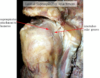
1. Supraspinatus: Performance cues- 1. Sitting: HBB (Hand behind back)- Adduction & medial rotation of the arm brings the tendon from frontal plane to sagittal plane. 2. Tendon emerge under the anterior edge of acromion 2. Infraspinatus: Performance cues- 1. Pr. Lying: prop up the patients on the elbows 2. Lateral rotation of the shoulder- ask the patient to hold to sides of the couch 3. Adduct the shoulder by shifting the affecting the elbow for 2-3 inches Locating the tendon- The insertion of Infraspinatus tendon is just below the lateral end of the spine of scapula 3. Subscapularis: Performance cues- 1. Sitting or ½ Lying: Therapist palpates for Bacipital tendon by repeatedly IR & ER of the arm. Just medial to the proximal tendon there is lesser tuberosity & the subscapularis (tendon feels as hard as the bone). The tendon extends down to the shaft of humerus distally. 2. Put patient’s hand on her thigh & apply the massage to specific points 3. Caution: Avoid...

Dr Alsears letter to me- From medicine to meditation

Image
From medicine to meditation This entire article is by Dr Al Sears. This letter was received by me on my E-mail address, which made a fantastic reading for me. Al Sears, MD 11903 Southern Blvd., Ste. 208 Royal Palm Beach, FL 33411 December 11, 2009 Dear satyajit, When I was in India I met “yogis” or meditation masters who had remarkable powers of concentration. Simply by focusing on their breath they could change their heart rate, raise their body temperature, and even walk on burning-hot coals. In your daily life you probably don’t have the need or opportunity to walk on hot coals, but the power to quiet your mind gives you more control over your health than any pill or prescription. Many of these yogis practice meditation. It’s easy enough to do on your own. There are even clinical studies that back it up. Studies show meditation: * Lowers blood pressure * Improves sleep * Reduces stress and anxiety * Improves immunity * Helps with pain management * Increases produc...

Issues in Academics: Writing a PhD Synopsis/Summary/Proposal

Issues in Academics: Writing a PhD Synopsis/Summary/Proposal

Effect of verbal cues on exercise performance

Image
Muscle timing and activation amplitude and movement can be modified with verbal cues (Lewis & Sahrmann ,2009). Lewis & Sahrmann tested the participants under 3 conditions: no cues, cues to contract the gluteal muscles, and cues to contract the hamstrings muscles. Hip and knee angle and electromyographic data from the gluteus maximus, medial hamstrings, and lateral hamstrings was measured while participants performed prone hip extension from 30 degrees of hip flexion to neutral. (see the figure above) This information is important for clinicians using prone hip extension as either an evaluation tool or a rehabilitation exercise.

Pregnancy changes them all- Stability, continence and breathing. The role of fascia in these changes.

Image
(Figure 1) Pregnancy & pelvic Girdle Pain (PGP): Pregnancy-related pelvic girdle pain (PRPGP) has a prevalence of approximately 45% during pregnancy and 20-25% in the early postpartum period. Most women become pain free in the first 12 weeks after delivery, however, 5-7% do not. Pregnancy & urinary incontinence (UI): Wilson, Herbison, Glazener, McGee & MacArthur (2002): found that 45% of women experienced UI at 7 years postpartum and that 27% who were initially incontinent in the early postpartum period regained continence, while 31% who were continent became incontinent. The abdominal canister (container) in pregnancy and delivery: Muscles and fascia of the lumbopelvic region play a significant role in a. musculoskeletal function b. continence and c. respiration It seems that there is an existence of an interlinked trio of lumbopelvic pain, incontinence and breathing disorders. Musculoskeletal loading & the interlinked trio: (see figure 1 above) Synergistic f...

Therapy with amitriptyline or physiotherapy is equally effective in fibromyalgia!!!

Image
You can learn about fibromyalgia from this blog site by typing “fibromyalgia” in the search box of this blog site. The search box is at top left of this page. In an rural set up Indian study, first of it’s kind, comparing the effect of amitriptyline or physiotherapy in improving outcome in patients of fibromyalgia over a period of six months, Joshi et al found they are equally effective. Usually fibromyalgia is treated by: antidepressants, analgesics, exercise, cognitive behavioral therapy. 1. Therapeutic Drugs used in fibromyalgia: amitryptiline and other tricyclic antidepressants, pregabalin, duloxetine and milnacipran. 2. Physiotherapy in fibromyalgia: Exercise in the form of cardiovascular training, strength training, aerobics, flexibility training, all can lead to improvement in fibromyalgia. Joshi employed a trained physiotherapist to conduct a uniform structured physical training and aerobic session for patients in physiotherapy treatment group. The patients were advised to perf...

Iliolumbar ligament under stress of slouching and the muscles that prevent it

Most biomechanical studies link the concepts of stooped postures and buckling instability of the spine under high compressive load. However everyday situations lumbar spine is subjected to small or neglectable compressive spinal load. Snijders et tried to find a mechanical cause of acute low back pain (LBP) in everyday situations. Hence their study in 2008 described strain on the iliolumbar ligaments (ILs) when slouching from standing upright. This study show that 1. Dynamic slouching, driven by upper body weight and rectus abdominis muscle force may produce failure load of the spinal column and the ILs. 2. There is a significant increase of IL elongation with rectus abdominis muscle force. 3. Contraction of erector spinae or multifidus muscle tension ease the ILs. 4. Sudden slouching of the upright trunk may create failure risk for the spine and ILs. This loading mode may be prevented by controlling loss of lumbar lordosis with erector spinae and multifidus muscle force. ...

The SI joint pain- Truth of provocative tests & SI joint blocks

Image
Because pain caused by sacroiliac joint dysfunction can mimic discogenic or radicular low back pain diagnosis of sacroiliac joint dysfunction is frequently overlooked, as common practice is to the link low back pain with protruding disc even when neurological signs are absent (1). The prevalence reported of SI joint caused pain is some where between 1. International Association for the Study of Pain (IASP) criteria demonstrated the prevalence of pain of sacroiliac joint origin in 19% to 30% of the patients suspected to have sacroiliac joint pain. 2. Hansen et al: the sacroiliac joint has been shown to be a source of pain in 10% to 27% of suspected cases with chronic low back pain utilizing controlled comparative local anesthetic blocks. 3. Rupert et al: prevalence of sacroiliac joint pain is estimated to range between 10% and 38% SI joint anatomy: The sacroiliac joint is a diarthrodial (freely moveable joint) synovial joint with abundant innervation and capability of being a ...

The LPSL- Lower back pain & deep gluteal pain considerations

Image
* LPSL = long posterior sacroiliac ligament In many patients with non-specific low back pain or peripartum pelvic pain, pain is experienced in the region in which the long dorsal sacroiliac ligament is located (3,1). 25 sides of the pelvis from 16 cadavers were studied by McGrath et al (2005) revealed that 1. LPSL is penetrated by the lateral branches of the dorsal sacral rami of predominantly S3 & and S2. Only in few cases LPSL is innervated by S4 and rarely by S1. 2. Some of the penetrating lateral branches give off nerve fibres that disappear within the LPSL ligament. These findings provide an anatomical basis for the notion that the LPSL is a potential pain generator in the posterior sacroiliac region. Same researcher McGrath et al in 2009 (2) reported few more interesting aspects of LPSL & it’s anatomical relationships 1. The LPSL was observed to have proximal and distal regions of osseous attachment. 2. Between these regions of attachment the middle LPSL was observed as a...

Viewing lumbar spine manual therapy from Lumbar paraspinal muscle EMG perspective

Introduction to approaches to investigating strategies in musculoskeletal conditions The approach is a comprehensive approach called physioanatomic approach that combines both anatomic approaches & physiological approach of investigation. Either the patient's pain pattern is categorized into a nonspecific pattern or into one of 4 recognizable pathway patterns i.e. radicular, dorsal ramus, polyneuropathy, and sympathetic. The goal of anatomic component of physioanatomic (both noninvasive and invasive) evaluation is to increase specificity by differentiating pain generators from asymptomatic underlying pathologic conditions. The physiologic component deals with function, reactivity & responsiveness of the designated or speculated pathologic tissues. There are 2 types of anatomic imaging approach i.e. invasive & noninvasive. Example of noninvasive anatomic imaging is computed tomography, single-photon emission-computed tomographic bone scan, and magnetic resonance im...

Basis of classification of LBA on EMG

Image
Persons with LBP often have reduced muscle strength and endurance, which may compromise the functional capacity of the spine and increase the likelihood of re-injury (Andersson, 1989). Historically, paraspinal muscle impairment has been quantitatively assessed by the use of dynamometers to supplement standard clinical assessment procedures (Roy SH, 1992). Assessments based on surface electromyographic (EMG) techniques have been proposed to overcome some of the problems inherent in the use of dynamometers for back muscle evaluation and classification (Merletti R, 1994). Recent approach of EMG variable is derived from the frequency rather than from signal amplitude of the signal. This is partly due to the fact that during a sustained contraction, the EMG signal propagates at a slower velocity and undergoes an alteration in shape associated with changes to the depolarization zone of the muscle membrane (De Luca CJ, 1985). These phenomena are referred to as "myoelectric manifestations...

The flexion relaxation phenomenon

I. Lumbar erector spinae flexion-relaxation phenomenon (FRP): A number of studies have shown differences in the FRP between patients with chronic low back pain and healthy individuals. Presence of the FRP during trunk flexion represents myoelectric silence (1). This leads to increased load sharing on passive structures and further these tissues have been found to fail under excessive loading conditions and are a source of low back pain. Persistent activation of the lumbar erector spinae musculature among patients with back pain may represent the body's attempt to stabilize injured or diseased spinal structures via reflexogenic ligamentomuscular activation thereby protecting them from further injury and avoiding pain (1). However, flexion relaxation phenomenon (FRP) is an interesting model to study the modulation of lumbar stability (2). II. Presence of fatigue of the erector spinae (ES) muscles modifies the FRP Descarreaux et al studied to identify the effect of erector spinae (ES)...

Long thoracic nerve injuries: new theories to consider

Image
The long thoracic nerve supplies the Serratus anterior, whose root value is (C5-C7) but the root from C7 may be absent. Anatomic path: The roots from C5 and C6 pierce the Scalenus medius, while the C7 root passes in front of the muscle. The nerve descends behind the brachial plexus and the axillary vessels, resting on the outer surface of the Serratus anterior. It extends along the side of the thorax to the lower border of that muscle, supplying filaments to each of its digitations (finger-like projections). Long thoracic nerve injuries in sports: Due to its long, relatively superficial course, it is susceptible to injury either through direct trauma or stretch. Injury has been reported in almost all sports, typically occurring from a blow to the ribs underneath an outstretched arm. Also injuries to the nerve can result from carrying heavy bags over the shoulder for a prolonged time. Symptoms are often minimal – if symptomatic, a posterior shoulder or scapular burning type of pain ma...

Scapular Dyskinesia

Image
Abnormal scapular motion is called scapular dyskinesis. Tennis players with scapular dyskinesia present a smaller subacromial space than non-athletes. Silva RT et al reported in Br J Sports Med (2008 Apr 8) that tennis players with scapular dyskinesia present a smaller subacromial space than control subjects. Additionally, when the shoulder was analyzed dynamically, moving from neutral abduction to 60 degrees of elevation, the tennis players with scapular dyskinesia presented a greater reduction in the subacromial space compared to unaffected athletes. Hence it seems logical that scapular dyskinesis is one causative factor in over-head athletic injuries. However the causal relationship between scapular dyskenesia & shoulder injuries has not been reported. Moreover reliable and valid clinical methods for detecting scapular dyskinesis are lacking. Let us discuss more on this issue on this subject & understand what exactly is scapular dyskenesia & how to test it. Following de...

Complete frozen shoulder lookout

Classification of FSS (Frozen shoulder syndrome) 1. Primary (Idiopathic) Frozen Shoulder 2. Secondary Frozen Shoulder 1. Systemic 1. Diabetes mellitus 2. hypothyroidism 3. hyperthyroidism 4. Hypoadrenalism 2. Extrinsic 1. Cardiopulomonary disease 2. Cervical Disc 3. CVA 4. humerus fractures 5. Parkinson's 3. Intrinsic 1. RTC Tendinitis 2. RTC Tears 3. Biceps tendinitis 4. Calcific tendinitis 5. AC arthritis *from Coumo, F. Diagnosis, Classification, and Management of the Stiff Shoulder. In: Disorders of the Shoulder: Diagnosis and Management. Iannotti, JP and Williams GR (eds). 1999 Description of pain in primary FSS (Frozen shoulder syndrome): The onset: After a period of pain, localized mostly in the shoulder and / or upper arm, begins the onset of severe limitation of movement of the glenohumeral joint in all directions. Cause: Movement limitation is caused by the retraction of the glenohumeral joint capsule and adhesions...

Be cautious while mobilizing stiff shoulder.

Current much acclaimed article of Vermeulen HM et al has changed the approach to frozen shoulder. End range mobilization is preferred over other techniques of mobilization. However, administering mobilization in shoulder is not out of danger. Drakos MC et al (The Hospital for Special Surgery, 535 E 70th St, New York, NY 10021, USA) reported “Shoulder dislocation after mobilization procedures for adhesive capsulitis”. This study is published in Orthopedics. 2008 Dec;31(12). No abstract or text available in PUBMED.

Leon Chaitow’s recommendation of manual therapy for parkinsonism

1. Antero-posterior and lateral mobilization of the thoracic and lumbar spine (patient seated). 2. Myofascial release of the thoracic spine (patient seated). 3. Atlanto-occiptal release (patient supine; not manipulation). 4. Mobilization of the cervical spine (patient supine). 5. Muscle-energy technique (MET) release of cervical muscles (patient supine). 6. General mobilization of the shoulder joints including use of MET (patient side-lying). 7. Mobilization of the forearms (patient supine). 8. Mobilization of the wrists (patient supine). 9. Mobilization of the SI joint (patient supine). 10. MET to the hip adductors (patient supine). 11. MET to psoas muscles (patient supine). 12. MET to hamstrings (patient supine). 13. Mobilization of the ankles (patient supine). 14. MET to the ankle in dorsi and plantar flexion (patient supine). Note: This sequence has to be performed in this order in 30 minutes.

Cadaver study of axial distraction mobilization of the glenohumeral joint support end range mobilization

The axial distraction mobilization techniques are frequently employed for treating patients with joint hypomobility. To know the biomechanical effects 3 different positions of glenohumeral abduction on a fresh cadaveric specimen ware chosen. They are 1. resting position 2. neutral position 3. end-range position Result indicated that displacement of the humeral head ware as follows: 1. largest in the resting position (27.38 mm) 2. followed by the neutral (22.01 mm) 3. and the end range position (9.34 mm). Greater gain in mobility was obtained in distraction at the end range position. During distraction mobilization, the force applied by the therapist and displacement of the humeral head depends on the joint position tested. These results also provide rationales for choosing end range distraction mobilization for improving joint mobility. Reference: Authors: Ar-Tyan Hsuab, Jing-Fang Chiuc, Jia Hao Changd