Posts

Mulligan’s positional fault corrections cause pain relief & mecanical corrections for long lasting effect!!!

Image
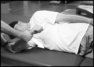
According to Vicenzino et al (2007) there are an increasing number of reports espousing the clinically beneficial effects of Mulligan's mobilization-with-movement (MWM) treatment techniques. The most frequent reported effect is that of an immediate and substantial pain reduction accompanied by improved function. Manual therapy effects on pain have been explained by many authors. The mechanism involved is thought to be an effect of mechanoreceptor response that affects the pain gait. Few others claim pain relief may be due to supra-spinal mechanisms based on opiate-like substance releases. However recent findings refer the supra-spinal mechanisms may not be involved especially spinal manual therapy-induced hypoalgesia. Naloxone antagonism and tolerance studies employ widely accepted tests for the identification of endogenous opioid-mediated pain control mechanisms. Paungmali et al (2004) reported that rapid initial hypoalgesia caused by Mulligan MWM was not antagonized by naloxone, ...

Viewing end plate from injury prospective

* This following information must be seen in the light of my previous 2 posts especially the last post. This class ends the series. 1. The vertebral body & the end plate: According to Prakash et al, many factors decide the integrity of the body of the vertebra. Gross design of the vertebral body is one of the most important adaptations for axial loading. The body of the vertebra is inter-segmental in origin, which results in dual vascular and nerve supply, both from superior and inferior aspects of the body. The vertebral body ossifies from 3 primary centers, one for centrum, which will form the major portion of body, and the other two for neural arches. The cartilaginous growth plate is mainly responsible for the longitudinal vertebral growth. 2. Intra-structural strength variation of end plates with specific reference to lumbar & sacral regions: Many studies indicate that some regions of the vertebral body may be stronger than others. Hence the failure strengths are different...

Schmorl's nodes

Image
Introduction Schmorl’s nodes are nothing but protrusions of disc material into the surface of the vertebral body i.e. intraosseous disk herniation. It is named after German pathologist Christian Georg Schmorl (1861-1932). Schmorl's node can be detected radiographically but it is imaged better by CT or MRI. MRI is not only useful in detecting the recently developed Schmorl's nodes but also in differentiating between symptomatic and asymptomatic Schmorl's nodes (4). With reference to Schmorl’s nodes discs in MRI are generally noted for size, location, margins, internal and surrounding T1/T2 signal, adjacent disc herniation or bulge, concentric ring, underlying fracture including malignancy, infection, or prior disc surgery. The migrating disc material of a Schmorl’s node when comes in contact with the marrow of vertebra it leads to inflammation. Many times protrusions are also associated with necrosis of the vertebral bone. Whether these protrusions and inflammation cause the...

Understanding Inter-vertebral disc & generation of pain from it

* The interveretbral disc is referred as disc in this following text Introduction: The intervertebral discs lie between the vertebral bodies, linking them together. The components of the disc are nucleus pulposus, annulus fibrosus and cartilagenous end-plates. The blood supply to the disc is only to the cartilagenous end-plates. The nerve supply is basically through the sinovertebral nerve. Biochemically, the important constituents of the disc are collagen fibers, elastin fibers and aggrecan. (3) The role that abnormalities play in the etiopathogenesis of different disorders is not always clear. Disorders may be caused by a genetic predisposition or a tissue response to an insult or altered mechanical environment. According to Roberts et al (2006), whatever the initial cause, a change in the morphology of the tissue is likely to alter the physiologic and mechanical functioning of the tissue (2) which is very important from all varieties (medicinal, physical therapy or surgical) of tre...

Kaltenborn: Foundation of treatment technique

The treatment goal: To restore joint play & normalize roll-gliding that occurs in normal active physiological movements 1. Resting & actual resting positions: resting positions is otherwise called maximum loose pack position. In this position the capsule is lax maximally hence can accommodate maximum most fluid. a. The term actual resting position is used for special circumstances where it is impossible, impractical or difficult to use maximum loose pack position. b. Positional fault: Traction to decrease pain is usually performed form resting position. If it is difficult to perform traction from the resting position then the actual resting position is chosen. If traction to reduce pain performed from actual resting position produces pain then a positional fault is said to exist. c. Once this is found first aim is to perform glide-mobilizations to correct the positional fault & it is obvious that one must find the direction of glide that is restricted &...

Learing the basic skills of manual therapy: Fundamentals of Maitland’s Mobilization

Image
Quotes from the legend: 1. The aim of examining movements is to find one or more comparable “signs” in an appropriate joint or joints. 2. Joint movements can never be classed as normal unless firm overpressure can be applied painlessly. 3. During examination and assessment pain should never be considered without relation to the range or vice versa. Performing the act: Different grades of movement are administered as oscillatory movements. Oscillatory movements have a positive & negative cycle. The positive portion of the cycle is one where impact of the force leads the accessory movement to sink into the tissues. Where as the negative portion of the cycle is one where, the therapist has to release the force to so that tissues can return to where it has started. Types of oscillatory movements: Oscillatory movements are administered in 1. Rhythmic manner or 2. With Irregular rhythm Under most circumstances the treatment used is regular & rhythmical. The informality...

Kaltenborn’s Approach to Joint Play Testing

Image
Key points 1. In contrast to Maitland, Kaltenborn developed his joint play testing with an emphasis on straight line, translatoric movement within a joint. 2. The use of CCR (concave convex rule) for joint play testing. 3. The examiner feels for abnormal resistance to motion with a particular emphasis on end-feel testing. 4. This testing is not truly oscillatory although it is often repeated several times using different speeds of movement. Points to focus • Bone rotation = Joint Roll-glide = Physiological movement • Bone translation = Joint gliding, Traction, Compression = Joint play Rotation = curved movement around an axis, Translation = straight-line movement Kaltenborn’s 3 Point Scale Kaltenborn developed a 3 point scale to describe the amount of movement and perceived resistance during manual joint testing and treatment. Slackness available can be referred as the mid point form feeling point of view. When slackness is taken-up stretching (Gr-III) starts. Before ...

VCS-vacuum cleft sign

1. The intravertebral vacuum cleft sign (VCS) is an uncommon radiological sign. 2. VCS is characterized by a radiolucent zone in the vertebral body. 3. This zone is composed of 95% nitrogen and small amounts of oxygen and carbon dioxide. 4. Cause: a. Post-traumatic ischemic necrosis (main cause) but other causes are (point b onwards) b. osteoporosis, c. corticosteroid therapy, d. diabetes, e. arteriosclerosis, f. alcoholism, g. multiple myeloma, h. bone metastasis and i. osteomyelitis. The broad diagnosis is made by AP X-ray. However DD encompasses CT scan and MRI. Reference: Sarli M et al; Osteoporos Int. 2005 Oct;16(10):1210-4. Epub 2005 Feb 25.

The Ultimate Frozen shoulder physical therapy recommendations

1. Pain reduction by Grade I cephalo-caudal glide recommended (Wardsworth CT; Physical therapy, Vol.66, dec.1986) 2. Mid-range mobilization II, II(-), II (+) (no specific effects claimed up to yet except as a progression of grade I) 3. End- range mobilization Increase in mobility & functional ability by grade III,IV ( Henricus MV et al, PHYS THER, Vol. 80, No. 12, December 2000, pp. 1204-1213 and PHYS THER,Vol. 86, No. 3, March 2006, pp. 355-368) 4. Mulligan’s mobilization with movement (MWM) correct scapulohumeral rhythm significantly better than end range mobilization (Yang JL et al Phys Ther. 2007 Oct;87(10):1307-15) 5. PNF techniques for shoulder (Joseph JG et al; J Orthop Sports Phys Ther; Vol.33, dec. 2003 ) - for scapulo-humeral alterations 6. PNF for upper trunk (My recommendation: Satyajit Mohanty, MSPT- not supported by research i.e. on clinical experience only) for scapulo-humeral alterations + upper trunk kyphosis

Challenging the CCR (concave-convex rule) mobilization convictions

Our peer’s classes on mobilization start with explanation of articular anatomy & McConnell joint classifications. It is followed by CCR with technique applications obeying the CCR. For shoulder mobilization it goes like this: 1. To regain ER – Anteriorly directed glides. 2. To regain ABD – Inferiorly directed glides 3. To regain IR – posteriorly directed glides Many times conventional wisdom is challenged when the research reports are contra posed. A paper JOSPT (2007) claimed that posteriorly directed joint mobilization technique was more effective than an anteriorly directed mobilization technique for improving external rotation ROM in subjects with adhesive capsulitis. However in groups comprised of both anterior directed glides compatible to CCR and posterior glides opposite to CCR recommendation had a significant decrease in pain. Reference: Johnson AJ et al; J Orthop Sports Phys Ther. 2007 Mar;37(3):88-99.

Which is better for clinical application of posterior capsule stretch- The Sleeper stretch or the Cross body stretch?

Image
Launder KG et al evaluated the acute effects of "sleeper stretches" on shoulder ROM. In their study sleeper stretches produced a statistically significant acute increase in posterior shoulder flexibility. However these authors explain that these acute changes in motion may not be clinically significant. Because of this recently expressed the belief that the sleeper stretch is better than the cross-body stretch to address glenohumeral posterior tightness because the scapula is stabilized McClure P et al compared changes in shoulder internal rotation range of motion (ROM), for 2 stretching exercises, the "cross-body stretch" and the "sleeper stretch," in individuals with posterior shoulder tightness. The sample consisted of 54 asymptomatic subjects (20 males, 34 females). The groups: 1. The control group (n=24) consisted of subjects with a between-shoulder difference in internal rotation ROM of less than 10 degrees. 2. Experimental groups ware those su...

Differences in major joint play testing grades

The pioneers of joint play testing The two principal pioneers of joint play testing in manual therapy are Geoffrey Maitland of Australia, and Freddy Kaltenborn of Norway. Both individuals developed techniques for the extremities and spine, and both developed different scales for describing the force and movement used during testing and treatment. Essential Differences between the Australian and the Nordic Approach During the development of his approach, Maitland was strongly influenced by the neurophysiologic principles relating to pain. Kaltenborn on the other hand was influenced by the joint based mechanical approaches advocated by Cyriax, Mennel, and Stodard. 1. Basic differences in approaches: Maitland’s approach (non-diagnostic approach): Maitland’s approach uses angular motions in the extremities when looking at joint play. Logically, angular movements of the long bones may be used to identify abnormal resistance due to muscle guarding, muscle tone, or articular restriction. How...

Grades of accessory movement

Grades of accessory movement This following article is about introducing the novice manual therapist to grades of applying passive accessory movement. Variables of Passive accessory movement administration 1. Direction 2. Grade 3. Speed & Rhythm 4. Duration of administration Aim of grading of passive accessory movements 1. To assess: Different abnormalities of spine & periphery presents with different amount of pain & resistance to passive movements. Our approach to palpation can not be the same to all of them as there may be different grades of tolerance to forces due to either mechanical faults or inflammation. a) Hence first of all force identified into different grades provides a rational explanation to approach tissue faults. b) It also helps to find out localized & referred pattern of pain (especially in spinal conditions). c) It further indicates tissue condition accessible at different depth of penetration (feel of the tissue). d) It als...

More about frozen shoulder (2): About End range mobilizations (High-Grade mobilization technique) & Early range mobilization (Low-Grade Mobilization

In year 2000 Henricus MV et al found End-range mobilization techniques with adhesive capsulitis of the shoulder increases glenohumeral mobility, but in the absence of a control group, they could not be sure what led to reduced impair as it is debated that natural course of the disease (FS) is self limiting. In 2006 Henricus MV et al found HGMT proved to be more effective than LGMT in the management of adhesive capsulitis of the shoulder; however, subjects improved significantly with both treatment strategies, and the differences were small. DESCRIPTION OF THE MOBILIZATION TECHNIQUES (AS ADVOCETED IN THE STUDIES OF HERNICUS MV ET AL) Each session must start with assessment of the ROM. All 3 affected physiologic movements of the glenohumeral joint are assessed passively. At each position of the shoulder, the end-feel of the movement are assessed in order to apply the mobilization techniques into the stiffness zone (HGMT group) or within the pain-free zone (LGMT group). 1. The treatmen...

Frozen shoulder as a algo-neuro-dystrophic process!

Waldburger et al (1992) of Switzerland subjected 50 cases of frozen shoulder across 3 Swiss medical centres to a radioisotope bone scan (99 mTc diphosphonate) study. They included 3 separate aetiological groups: post-traumatic (40%), neurological (14%) and idiopathic (46%). The findings are interesting: a. The so-called idiopathic frozen shoulder showed a scapulo-humeral increase in radioisotope uptake in several areas (in 82% of cases) without involvement of the ipsilateral carpus. b. Clinically, the neurological type was associated with a shoulder-hand syndrome with positive bone scan of the shoulder and the wrist in all cases. c. The post-traumatic type showed a diffuse (in 50% of the cases) or at several circumscribed areas (also in 50%) increase in radioisotope uptake in the shoulder. In 45% of the post-traumatic type, there was also a shoulder-hand syndrome with uptake in the wrist also. These findings made the resesrchers to intervene the FS subjects with calcitonin adm...

More on frozen shoulder & techniques of frozen shoulder mobilization (1)

Types of FS: Idiopathic FS- When no secondary causes are attributed or no cause can be assigned to the onset. Secondary FS: FS as a result of diabetes, cardiac problems, stroke, rheumatoid arthritis, or trauma. Phases of FS: Reeves documented 3 phases with which to address the progression of FSS: the pain phase, the stiffness phase, and the recovery phase. To regain the normal extensibility of the shoulder capsule and tight soft tissues, passive stretching contrast to active stretching of the shoulder capsule and soft tissues by means of mobilization techniques has been recommended. The in-vogue techniques encompasses 1. Midrange mobilization (MRM)- recommended by Maitland 2. End-range mobilization (ERM)- recommended by Kaltenborn 3. Mobilization with movement techniques (MWMs)- recommended by Mulligan However these above recommended techniques for FS base are not based on research they are rather suggestions. Yang et al investigated the effect of mobilization treatment and to...

Lumbo-coxa contribution to forward bending in symptomatic & asymptomatic LBA cases.

I. Asymmetry 3-dimensional motion patterns in Chr. LBA Chronic LBP patients exhibited motion patterns altered from those of the normal population. 3D motion analyses reveal 3 distinct patterns of motion observed in each principal direction of movement in LBA cases. They are reflected as: 1. Differences in the extension-flexion ratio (asymmetry between flexion and extension). 2. Lateral bending asymmetry and 3. Differences in coupled axial rotation-lateral bending ratio. II. Lumbar-hip flexion motion in LBA A. Hip movements in normal healthy patients without LBA during forward bending of the lumbar spine: 1. Earlier reported values for lumbar spine motion during forward bending vary from 23.9 degrees to 60 degrees and hip motion during forward bending ranges from 26 degrees to 66 degrees. 2. Esola & colleagues found mean total forward bending to be 111 degrees: 41.6 degrees from the lumbar spine and 69.4 degrees from the hips. B. The lumbo-coxa contribution to forward bendi...

Spine Asymmetry & LBA (specific focus on lumbo-pelvic spine)

Introduction: Subtle pelvic asymmetry (exhibited as either lateral pelvic tilt or iliac rotational asymmetry), which is common among normal individuals, has not been convincingly linked to abnormalities in back. Given the difficulty in diagnosing most LBA, a classification using pelvic asymmetry and patterns of movement are helpful in establishing a rational treatment plan (2). I. Impact of pelvic asymmetry in work-stations: Structural and functional asymmetries are factors that may be considered in the seating design and work environment (1). A study consisting of 59 subjects revealed that significant: (1) Correlations between pelvic asymmetry and asymmetric trunk motion performed in sitting. (2) Differences between the LBP and control groups in patterns of trunk motion performed in a sitting posture. (3) Differences between kinematics of motions performed in sitting versus standing postures. This study concluded that in LBA cases pelvic asymmetry and altered trunk motion in...

Assessment of the spine asymmetry

Modern manual therapy techniques focus on asymmetry in anatomy & mobility and lateralization. Wylick HV et al recommend a 3 Minuit test battery by 8 tests. How ever following are 10 tests to know the asymmetry in spine: 1. Dominant eye 2. Phoria of eye 3. Scooping 4. Step forward 5. Finger crossing 6. Step on platform 7. Axis rotation 8. Tailor’s position 9. Step backward 10. Arm crossing

Taxonomy to describe treatments for musculoskeletal pain

According to Rubik & colleagues (1994) massage therapy, the manual manipulation of soft body tissues to enhance health and well-being, is one of the oldest forms of medicine known to mankind and has been practiced worldwide since ancient times. Today, more than 80 different forms of massage have been identified, many developed in the last 30 years. Lack of consistent terminology for describing the treatments given by therapists are felt world wide. Sherman & colleagues developed taxonomy to describe therapist guided module delivery for patients with musculoskeletal pain. Due to this work a new classification system evolved. Using this, practitioners using different styles of extramural medicine (manual medicine technique) can describe the techniques they employ using consistent terminology. About the study: A review of the literature for treatment musculoskeletal pain was done for creating the taxonomy & neck pain was the matter subjected to further studies. The results war...